Watch


Why the navel, and why first
Pain or hardness around the navel reports disturbed blood flow
Every therapy must answer one question before it can begin: where do I look? A physician answers it with a history, a stethoscope and a laboratory. This therapy answers it with the belly, in the first two minutes — and the reason is not sentimental. It rests on an observation made over decades, confirmed on lakhs of patients, checkable by anybody on the next ten people they examine.
The observation the whole map rests on
Specific points around the navel, in front, exhibit pain or hardness — and the same points are found tender in all patients sharing a particular symptom or group of symptoms.
More broadly: any disease, whatever its individual symptoms, is generally accompanied by pain or hardness around the navel and the hip, at one or more of eighteen specific points.
And pain in an area indicates some disturbance in the blood flow to that area, and to the organ or organs associated with it.
Read the last line backwards, because that half is the working half: if the pain at a place is relieved, the blood supply to that place has been restored. Removal or reduction of one or more of those pains produces an immediate sense of well-being, which the patient herself confirms without being asked.
Two words in that teaching do quiet work. Hardness — you are feeling not only for a wince but for a place where the tissue has lost its give, where the belly wall meets your finger like a half-inflated ball instead of a cushion. A patient can be stoical about pain and cannot be stoical about hardness, because hardness is not an opinion. Group of symptoms — the point does not belong to a disease but to a pattern, which is why two people with different disease names on their cards can be tender at the same point and need the same treatment, and why one person is tender at different points on Monday and Thursday.
Why the navel in particular? Because of where the body was assembled from. Every one of us was built outward from that point; in the womb it is the door through which everything arrives, and the organs of the abdomen take their places at fixed distances from it, each at its own settled remove from all the others. Nothing in the abdomen has spare room. The organs are packed with such skill that no empty space is left over.
That packing is why the region is worth reading. For an organ to work smoothly it must go on receiving, twenty-four hours a day and without interruption, blood through the finest vessels and messages along the nerves — and those vessels and nerve bundles are exceedingly fine, running between the organs through the gaps. If through some unusual movement the internal gaps alter, pressure can fall on a capillary or a nerve bundle. The obstruction that follows need not be dramatic. First the stomach is upset; then, slowly, the balance of the whole body is disturbed.
How the map was built
Decades of pressing the same places, and the code names that followed
Nobody designed this chart; it accumulated, by the least glamorous method available. Press the same places on very large numbers of people, write down what you find, and wait for the pattern to declare itself. Over five decades and something approaching a million patients, one fact kept repeating: people who shared a complaint were tender in the same square inch, and that square inch was reliable enough to be marked on a drawing and taught to a stranger. From it came the instrument this article teaches — the chart of pain points, the main basis of diagnosis in this therapy and the page a student returns to more than any other.
The points carry code names — symbolic short names, generally indicating the principal organ or symptom of that region. Generally, and not invariably: the qualification is part of the teaching, and a student who forgets it will make the commonest mistake in .
Four further points lie near the hip, above the front and the back of the hip bone, and these are indicative of certain vitamin deficiencies, carrying the same code names as the deficiencies they answer.
The front chart, complete
Every front point — where it lies, what is beneath, why the name
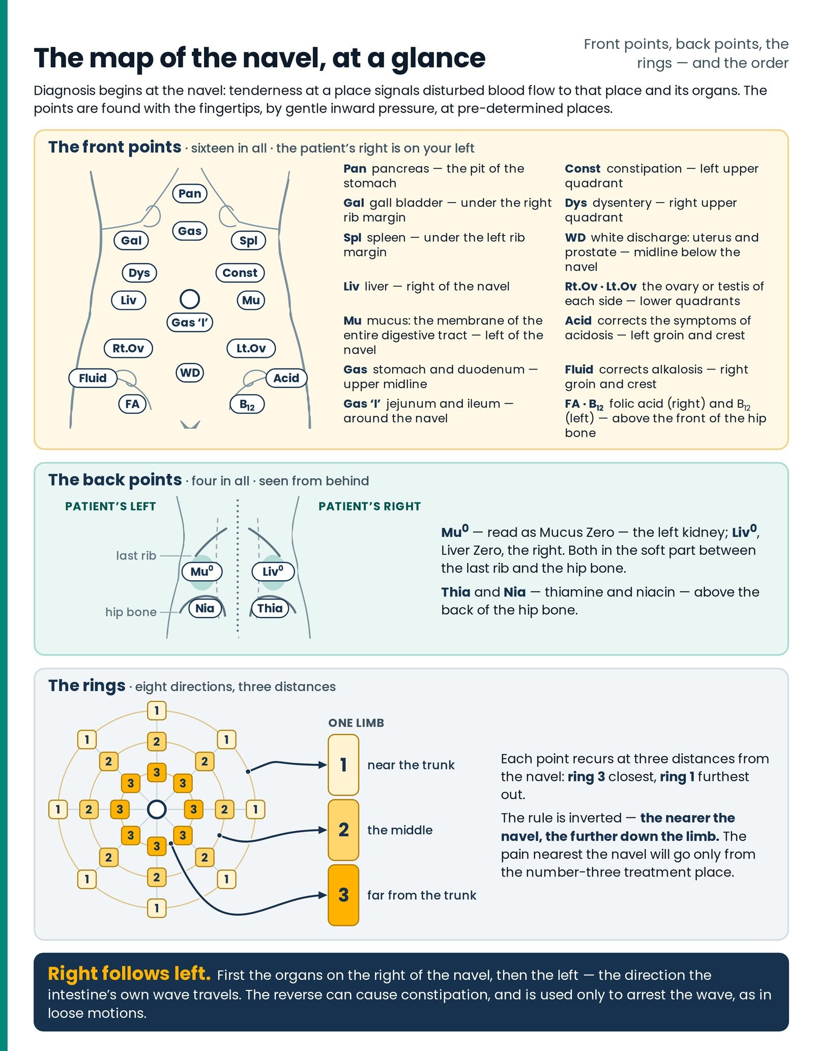
The front chart is a line drawing of the trunk from neck to upper thighs, with the rib margins sketched in, the crests of the hip bones, the creases of the groin, and a small open circle for the navel. Three structures are marked on the midline above the field of points — the thyroid at the base of the neck, the thymus behind the upper breastbone, the pancreas as a small oval at the very bottom of it. Around and below the navel lies a field of small numbered boxes carrying 1, 2 or 3, and eighteen labelled call-outs run down the two margins of the page to reach them. (Figure 6.1)
Here is the whole front list. Learn the table first and the reasoning afterwards; the reasoning will not stay in a mind that has no pegs to hang it on. (Figure 6.8)
Table 1 — The pain points of the front chart
| Code | Full name — organ or symptom | Where it lies, by feel |
|---|---|---|
| Pan | Pancreas | The pit of the stomach — the soft dip at the bottom of the breastbone, where the two rib margins meet in a shallow V |
| Gal | Gall bladder | Under the right rib margin, where the outer edge of the straight belly muscle crosses it Right |
| Spl | Spleen | Under the left rib margin — the mirror of Gal Left |
| Liv | Liver | To the right of the navel, level with it, in the middle ring Right |
| Mu | Mucus — the mucous membrane of the entire gastrointestinal tract | To the left of the navel, level with it, in the middle ring Left |
| Gas | Stomach and duodenum | The midline of the upper belly, below the breastbone, above the navel |
| Gas 'I' ("Gas eye") | Jejunum and ileum | Immediately around the navel |
| Const | Constipation — kabjī | Left upper quadrant, middle ring, between navel and left rib margin Left |
| Dys | Dysentery — loose motions | Right upper quadrant, middle ring, between navel and right rib margin Right |
| WD | White discharge — uterus and prostate gland | The midline, below the navel |
| Rt.Ov | Right ovary / right testis | Right lower quadrant, midway between the navel and the bony front point of the right hip Right |
| Lt.Ov | Left ovary / left testis | Left lower quadrant — the mirror of Rt.Ov Left |
| Acid | Corrects the symptoms of acidosis | Over the left groin and the crest of the left hip bone Left |
| Fluid ("Alkali or Fluid") | Corrects the symptoms of alkalosis | Over the right groin and the crest of the right hip bone Right |
| FA / Folic | Folic acid deficiency | Above the front of the right hip bone, at the groin and crest Right |
| B₁₂ | Vitamin B₁₂ deficiency | Above the front of the left hip bone, at the groin and crest Left |
How the labels sit around the page. The chart is an anterior view, so the patient's right is on the reader's left. Down the reader's left margin — the patient's right — run GALL, DYS, GAS, LIVER, RT.OV., and at the foot THIAMIN, FOLIC and ALKALI OR FLUID. Down the reader's right margin — the patient's left — run SPLEEN, CONST, MUCUS, NAVEL, LT.OV., and at the foot NIACIN, B₁₂ and ACID.
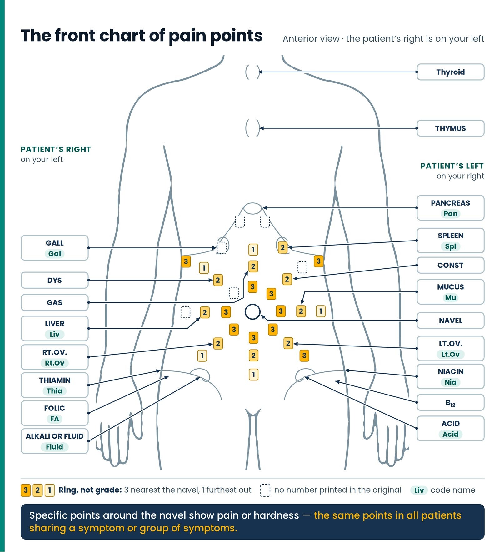
Now take them one at a time: how to find each on a living body, what is under your finger, and why it carries that name.
Pan — the pancreas point
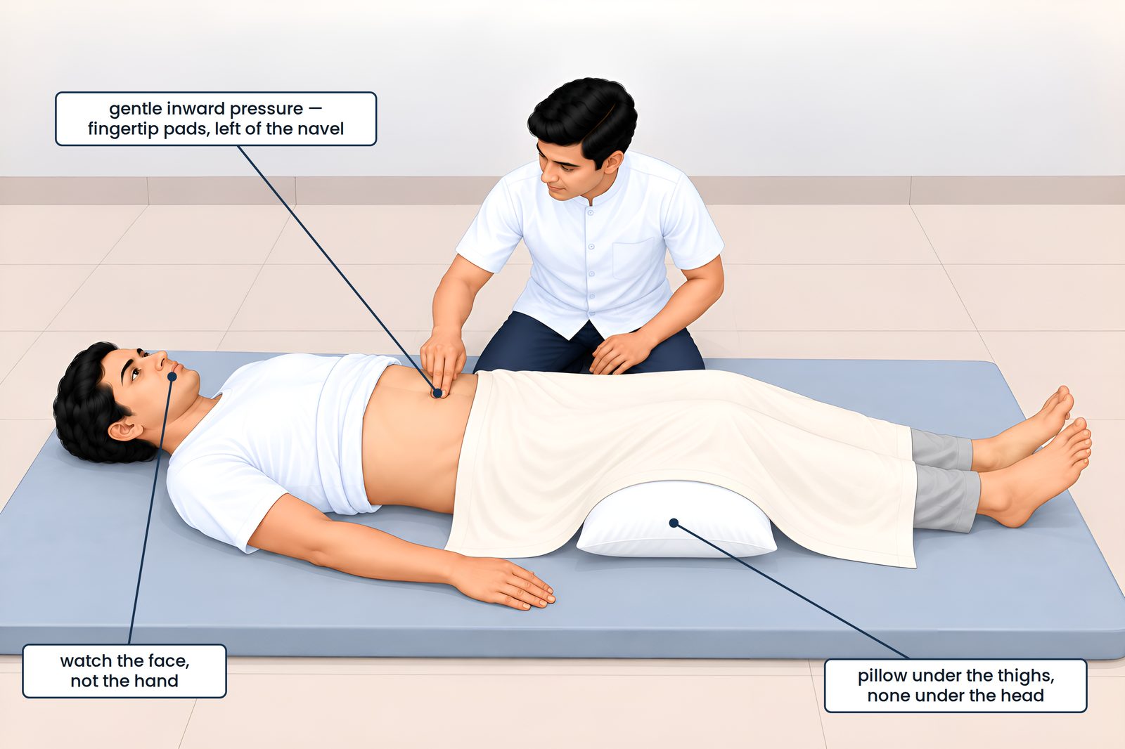
Finding it. Run two fingers up the midline from the navel until the belly wall ends and bone begins. Where the rib margins meet, at the bottom tip of the breastbone, there is a small soft dip — the spot a person rubs with the heel of the hand and calls the pit of my stomach.
What is beneath. The midline seam where the two straight muscles meet edge to edge; behind it the left lobe of the liver and the upper stomach; and crossing the back wall at about the first lumbar bone, lying across the great artery like a wet cloth over a rope, the body of the pancreas.
Why the name. In diseases connected with the pancreas, pain or stiffness is found around the navel at just this place. The chart marks the pancreas here as a small oval — the one front point where the named organ genuinely lies more or less behind the mark.
Gal — the gall bladder point
Finding it. Lay your hand flat under the patient's right rib margin and find the outer edge of the straight belly muscle — ask her to lift her head an inch and it stands up under your fingers, then let her lie back. Where that edge crosses the rib margin, at the tip of the ninth rib cartilage, is the point. Right
What is beneath. The rounded end of the gall bladder, poking out from under the liver's edge; behind and above it the right lobe of the liver, beside it the bend of the large bowel, just deeper the first part of the duodenum.
Why the name. In gall bladder trouble, pain or stiffness is found in just this part. It is also the exact spot a surgeon presses when he suspects an inflamed gall bladder — two traditions arriving at one square inch from opposite directions, worth saying to a doctor who asks.
Spl — the spleen point
Finding it. The mirror image of Gal, under the left rib margin. Left
What is beneath. Not the spleen — that sits further back and higher, under ribs nine to eleven, and in health cannot be felt at all. Under your finger here are the stomach, the bend of the large bowel under the spleen, and the great apron of fat over the intestines.
Why the name. Because in diseases connected with the spleen, pain or stiffness is found here — an observation about tenderness, not a claim that you are touching a spleen.
Liv — the liver point
Finding it. Two to three finger-breadths to the right of the navel, level with it, in the middle ring. Right
What is beneath. The straight belly muscle in its sheath; behind it, in a thin patient, the lower edge of the right lobe of the liver; below and behind, the ascending colon, with small-bowel loops between.
Why the name. In liver disease, pain or stiffness is found here. Remember its side — right of the navel — because it is half of the memory device naming the kidney points at the back.
Mu — the mucus point
Finding it. The mirror of Liv, the same distance to the left of the navel and level with it. Left
What is beneath. Straight belly muscle; behind it the descending colon, and coils of jejunum and ileum.
Why the name. Whenever slimy mucus — āṃva — is coming in the motions, there is pain at this point. And a second observation pushed the name outward from the bowel to the whole body: if a person has a blocked nose or a dry mouth, a particular treatment is given on the right thigh and hand, and moisture then comes into the nose and the mouth. This has been proved on many patients. From it Guruji says that our treatment provokes the mucous membrane — and it is for that reason that the treatment was named Mucus.
So the hash mark on the chart is not a footnote to skip. Mu does not stand for mucus in the bowel; it stands for the mucous membrane of the entire gastrointestinal tract — one continuous wet lining from the lip to the far end, behaving as the lining of nose and mouth behaves.
Gas — the stomach and duodenum point
Finding it. The midline of the upper belly, between navel and breastbone, in the middle ring; the chart reaches it on a long leader.
What is beneath. The body and outlet of the stomach; the first and second parts of the duodenum curving right; below them, running across, the transverse colon.
Why the name. Not "gas" in the sense a patient uses the word. Gas codes for the stomach and duodenum — the first stretch of the tube, where food is broken down and handed on. A student who reads Gas as wind will misread half the formulary.
Gas 'I' — the jejunum and ileum point
Finding it. Immediately around the navel itself.
What is beneath. The small intestine — coils of jejunum and ileum on their fan of mesentery, mobile enough to slide under your finger.
Why the name. Gas 'I' is the small-bowel point as Gas is the gastro-duodenal one; read the I as intestine and the pair keeps itself straight. This is where absorption happens — where the raw materials every gland depends on either get into the blood or fail to.
Const — the constipation point
Finding it. The left upper quadrant, middle ring, on a line from the navel towards the left rib margin. Left
What is beneath. The descending colon and the bend of the bowel under the spleen — where formed stool waits before it moves on. Press it in a constipated patient and you may feel not only tenderness but a firm, sausage-like fullness that does not yield.
Why the name. One of the two points named for a symptom rather than an organ, asterisked on the chart to say so. In constipation there is sharp pain at just this point.
Dys — the dysentery point
Finding it. The right upper quadrant, in the middle ring — the mirror of Const. Right
What is beneath. The ascending colon and the bend of the bowel under the liver; below them the caecum and the last stretch of small intestine.
Why the name. The other symptom-named point: in loose motions there is sharp pain at just this point. The side is part of the instruction — the finding that licenses the Dys treatment is pain in the upper part to the right of the navel. Right
WD — the white discharge point
Finding it. The midline below the navel, between navel and pubic bone.
What is beneath. The midline seam; behind it the dome of the bladder when full — one good reason a patient passes urine before a session — the uterus resting above the bladder in a woman, and loops of sigmoid colon.
Why the name. Whenever women have the problem of white discharge, pain or stiffness is found at just this place below the navel; and as soon as the treatment named WD is given, both the pain and the discharge come to an end. The code covers the corresponding organ in either sex: uterus and prostate gland.
Rt.Ov and Lt.Ov — the gonad points
Finding them. Put a fingertip on the navel and another on the bony point at the front of the hip — the knob you feel with your hands on your hips. Halfway along that line, in the lower quadrant, is the point: Rt.Ov on the right Right , Lt.Ov on the left Left .
What is beneath. The hollow of the iliac fossa; the great vessels running down to the leg; the tube from the kidney crossing the brim of the pelvis. The ovary itself lies deep on the side wall of the pelvis and cannot be reached by a finger on the belly, and in a man no gonad lies here at all.
Why the name. In diseases connected with these organs, pain or stiffness is found at just these places. The codes cover left ovary or testis and right ovary or testis — one point, either sex.
Why two separate codes and not one pair. Because the two are not interchangeable. Wherever the body has two glands of a kind, their work is nevertheless not the same: more oestrogen comes from the left ovary, more progesterone from the right — which is why the treatments for menstrual disorder are given on one side for the first fortnight after the menses and on the other for the second.
Acid and Fluid — the two balance points
Finding them. Not around the navel but down at the hip. Acid lies over the left groin and the crest of the left hip bone Left ; Fluid — labelled on the chart as ALKALI OR FLUID — over the right groin and crest Right .
What is beneath. The crest of the hip bone and the muscle sheets taking origin from it; the ligament of the groin and the canal running along it; the two nerves supplying this strip of skin; and just below the ligament, the great vessels passing into the thigh.
Why the names — and the trap in them. These two are named neither for an organ nor for a symptom but for what pressing them does. Acid corrects the symptoms of acidosis; Fluid corrects the symptoms of alkalosis. So Acid is not the acidosis point — it is the anti-acidosis point, and a student who reads it the other way will give the wrong one of the pair. Note their sides and hold them until The Two Kingdoms: Acid sits on the left, and pains on the left of the navel generally keep company with the symptoms of acidosis; Fluid sits on the right, with alkalosis.
FA and B₁₂ — the two vitamin points of the front
Finding them. Near the hip, above the front of the hip bone, at the groin and the crest — FA, folic acid, on the patient's right Right ; B₁₂ on the left Left . Pain at these points is connected with the symptoms of deficiency of just these vitamins, and each carries the name of the deficiency it answers. They are two of the four pain points near the hip; the other two lie above the back of the hip bone and belong to the back list.
The four vitamin points, front and back
FA, B₁₂, Thia and Nia on the rim of the hip bone — prompts to test
Take the four together: they behave as a family, and they are the points most likely to send a patient into a laboratory.
Table 2 — The four vitamin points near the hip
| Code | Deficiency it is linked with | Where it lies |
|---|---|---|
| FA / Folic | Folic acid | Above the front of the hip bone, right groin and crest Right |
| B₁₂ | Vitamin B₁₂ | Above the front of the hip bone, left groin and crest Left |
| Thia | Thiamine — vitamin B₁ | Above the back of the hip bone, on the right Right |
| Nia | Niacin — vitamin B₃ | Above the back of the hip bone, on the left Left |
Two things about this table must be taken exactly as they stand.
First, the code lists place Thia and Nia on the back — above the back of the hip bone — while the front chart carries their labels too, printed at the foot of the page with their leaders converging on the groin and the crest: THIAMIN on the patient's right beside FOLIC and ALKALI OR FLUID, NIACIN on the patient's left beside B₁₂ and ACID. The crest is a rim, not a point, and it has a front and a back; all four vitamin points live on that rim.
Second, apart from vitamin K and folic acid the vitamins are not made in the body — they must come from food. Certain pains behind the back are linked with certain vitamin deficiencies, and as the digestive system is set right and the pain is drawn out of those points, the symptoms of the deficiency begin to reduce. The route is absorption, not manufacture.
The back chart — Mu° and Liv°
The kidney points in the soft hollow between last rib and hip bone
Turn the patient, or reach round to the flank, and the map continues. The back list is short, and it is the half of the chart a therapist uses first.
Table 3 — The pain points of the back
| Code | Read aloud | Principal organ or related symptom | Where it lies |
|---|---|---|---|
| Mu° | Mucus zero | Left kidney | The soft part in the middle, between the last rib and the hip bone, on the left Left |
| Liv° | Liver zero | Right kidney | Exactly the same place on the right Right |
| Thia | Linked to thiamine deficiency symptoms | Above the back of the hip bone Right | |
| Nia | Linked to niacin deficiency symptoms | Above the back of the hip bone Left |
Finding Mu° and Liv° by feel
You find this point with your hand and not with a ruler, so it is taught by feel.
Why that spot is soft, and what sits behind it
The softness is not fat and not an accident. It is the one window into the back of the abdomen that no bone covers.
Going in: skin and fat; then the strong sheet of fascia across the low back into which the great back muscle inserts; then, in the small triangular gap between that muscle, the flat muscle of the flank and the rim of the hip bone, a place where the wall is genuinely thin — which is why your finger reports soft. Deeper, the square muscle running from hip rim to last rib, and beside it the great hip flexor; crossing between the layers, the nerves carrying sensation from this strip of skin — which is why tenderness here can be muscular or nerve-borne rather than anything to do with a kidney. Then the space behind the abdominal cavity proper: a membrane like a paper bag packed with fat, and in that fat the kidney, the right one about half a bone lower than the left because the liver takes the room above it. Capping each kidney like a small hat, the adrenal gland.
The two reasons for the code names
Now the naming, which is one of the most humane pieces of thinking in the whole therapy.
Why the kidneys are called Mu° and Liv°
The first reason is to spare the patient alarm.
Stimulation of the kidney is a necessary part of a great many treatments, so the word would otherwise be spoken constantly — across a treatment room between therapists, and in writing on a card the patient can read. But kidney is a much-maligned word: in many patients it conjures dialysis, hospital wards, an operation, and in an anxious one the mere utterance of it can send the blood pressure up. To save patients from such baseless fears, the kidneys were given the nicknames Mu° and Liv°.
The second reason is memory. The Liv pain point lies on the right of the navel, and the right kidney lies immediately behind it — so the right kidney is called Liv°. Right The Mu pain point lies on the left, and the left kidney sits just behind it — so the left kidney is aptly called Mu°. Left
Learn the two front points and you have the two back points for nothing. That is the whole design.
And the ring itself carries information. It is not decoration and it does not mean zero.
What the small ring means
The place and the manner of giving treatment for Liv and Liv°, and for Mu and Mu°, are the same. But there are two changes in the method, and the ring records both.
One — one place instead of three. Where Liv and Mu are given from three places in turn along the limb, the ° points are given at a single point; to indicate that, the sign ° is used.
Two — the hand point moves up to the shoulder. The site of the point on the arm is up on the shoulder, shown by writing the ring as a superscript: the mark sits high, above the line, to remind the therapist that the contact goes high, near the shoulder joint, and not at the elbow where Liv and Mu take it.
One mark, at once a warning, a memory aid and a picture of the instruction.
At every step you can see the thoughtfulness behind this: with two logically worded abbreviations, therapist and patient were both put at ease in one stroke.
Two rules keep that kindness honest. A code may conceal a word; it may never conceal a finding — if you find something that ought to send a patient to a doctor, the nickname stops there and you say the real thing plainly, to the patient and on the paper he carries. And when a patient asks what it means, tell him: "It is our shorthand for the kidney. We use a short name because the word frightens people, and there is nothing here to be frightened of." A patient who discovers later that you had a private word for his kidney will not remember it as tact. ▲ Take care
What a finding at Mu° or Liv° means belongs to the next stage of your training. Tenderness at these two is the first thing checked in a Neurotherapy examination, and it sorts the patient into one of two great families — the acid direction or the alkaline — from which a very large part of the plan follows. That is taught completely in The Two Kingdoms, "The Two Kingdoms", with the two-column symptom picture, the dilution rule and the asymmetry of the two kidneys. Learn the anatomy and the naming here; learn the reading there.
The back pain chart
Twelve sites where the back reports an internal organ’s trouble
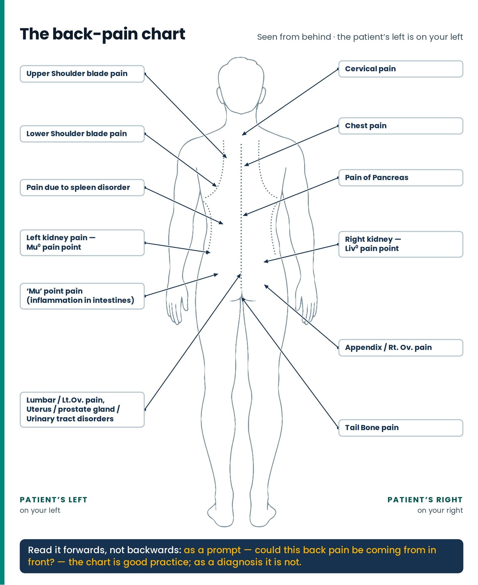
The second full-page chart is a whole-body outline seen from behind, head to heels, the spine a dotted line down the middle, the shoulder blades and lower rib margins dotted curves either side. Twelve labelled call-outs run around it, and together they make a compact statement of something the body does constantly and confusingly: it reports the trouble of an internal organ as an ache in the back wall. (Figure 6.2)
One drafting point must not catch you. The drawing is a view from behind, so the patient's left is on the reader's right — but the twelve call-out boxes have been placed for the sake of the page, the left-sided labels sitting on the reader's left and the right-sided ones on his right, the arrangement that would be correct facing the patient's front. A student who learns the chart as a picture will reverse every side on it the first time he stands behind a patient. Locate these points by anatomy, never by the side of the page: spleen and Mu° on the patient's left; liver, Liv° and appendix on the patient's right. Left Right
Table 4 — The back pain chart, label by label
| Label on the chart | Where the arrow lands | What lies at that level |
|---|---|---|
| Cervical pain | Nape, base of the neck, midline | The seven neck bones and the long muscles either side |
| Upper Shoulder blade pain | Upper scapular region, inside the shoulder blade | Upper trapezius, the muscle that lifts the shoulder blade, the small rhomboid, over ribs two to four |
| Lower Shoulder blade pain | At the bottom corner of the shoulder blade | Large rhomboid, lower trapezius, ribs six to eight; the base of the lung deep to it |
| Chest pain | Upper back, beside the spine | The first six thoracic beads, the long back muscles, and the joints where each rib meets the spine |
| Pain due to spleen disorder | Lower thoracic, below the shoulder blade | Ribs nine to eleven and the fascia of the low back; deep to them, spleen left, liver right |
| Pain of Pancreas | Mid-thoracic beside the spine | Long back muscles, ribs ten to twelve; the pancreas lies at the front of this level |
| Left kidney pain — Mu° pain point | The flank, between the twelfth rib and the rim of the hip bone Left | The square muscle of the low back, the hip flexor, the fat around the kidney, and the kidney |
| Right kidney — Liv° pain point | The mirror-image flank Right | The same, the right kidney about half a bone lower because of the liver above it |
| Mu point pain — inflammation in intestines | Lower lumbar, out from the spine | Long back muscles and fascia; deep and in front, the descending colon and small-bowel loops |
| Lumbar / Lt.Ov pain — uterus, prostate gland, urinary tract disorders | Lower lumbar and upper sacral region | The junction of low back and sacrum, the joints between sacrum and hip bones; deep in front, the tubes from the kidneys, the bladder, the uterus or prostate |
| Appendix / Rt.Ov pain | Lower lumbar to the sacro-iliac joint, opposite side | That joint and the bony knob behind it; deep in front, the caecum and appendix, or the pocket in which the ovary sits |
| Tail Bone pain | Coccyx, top of the cleft of the buttocks | The tail bone and its joint with the sacrum; the muscles of the pelvic floor taking their attachment there |
The chart is intelligent about a real thing. A pancreas in trouble really does bore through to the mid-back and ease when the patient leans forward; a kidney in trouble really is felt in the loin between last rib and hip rim, running towards the groin as a stone travels; trouble in the womb or prostate really is felt low in the back and over the sacrum. But read it forwards, not backwards. Most back pain at every one of these levels is muscle and posture, and an ache between the shoulder blades is far more often a woman who sews for nine hours than a pancreas. As a prompt — could this mid-back pain be coming from in front? — the chart is good practice; as a diagnosis it is not.
The ring rule — how to read the numbers on the chart
Rings, not grades: the nearer the navel, the further down the limb
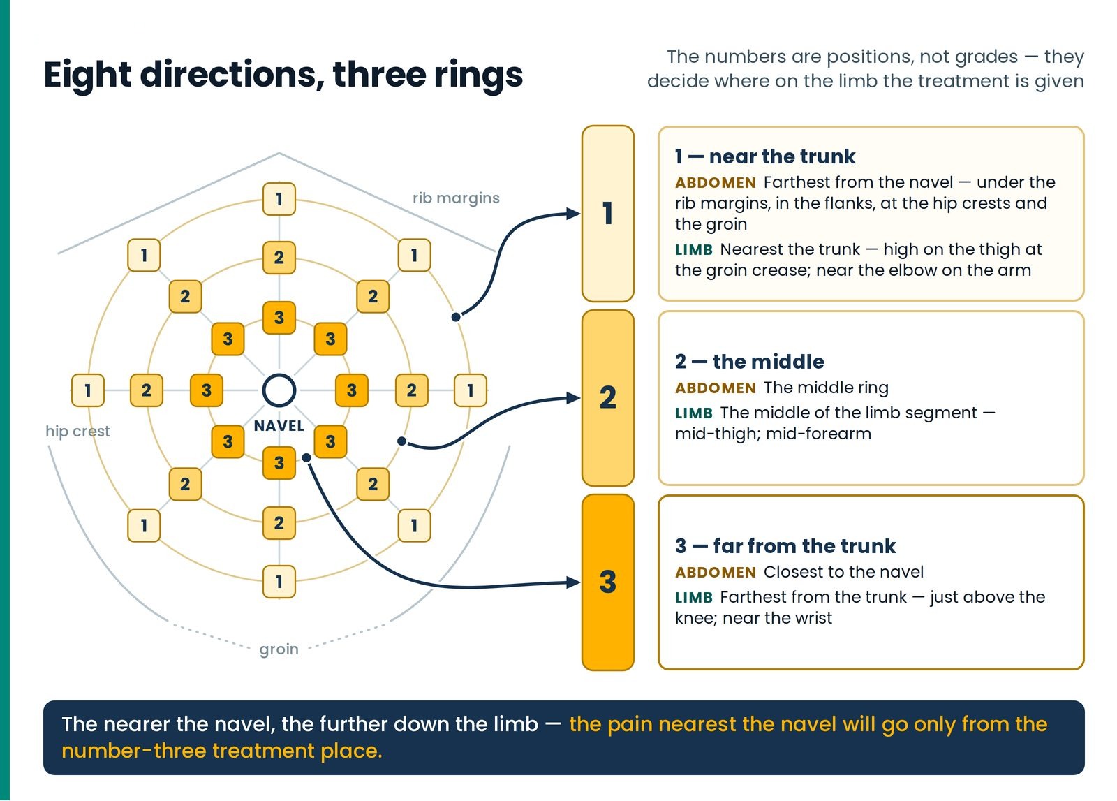
Now the part of the chart beginners skip and should learn before any point name: those small boxes carrying 1, 2 and 3. They are not grades of severity — a 3 does not mean worse than a 2 — but coordinates, and they decide where on the limb the treatment is given. (Figure 6.3)
In the eight directions around the navel — the four cardinal directions and the four diagonals — numbers such as one, two and three have been given along with each name.
The eight directions and the three rings
These numbers are connected with the order of our treatment. They tell which pain will go by giving treatment where — that is, the pain of a number-one place will go from the number-one treatment place.
Along every one of the eight directions, the same named point recurs at three distances from the navel:
the 3s cluster immediately around the navel;
the 2s form a middle ring;
the 1s lie furthest out — towards the rib margins, the flanks, the crests of the hip bones and the groin.
And the general rule that binds it together:
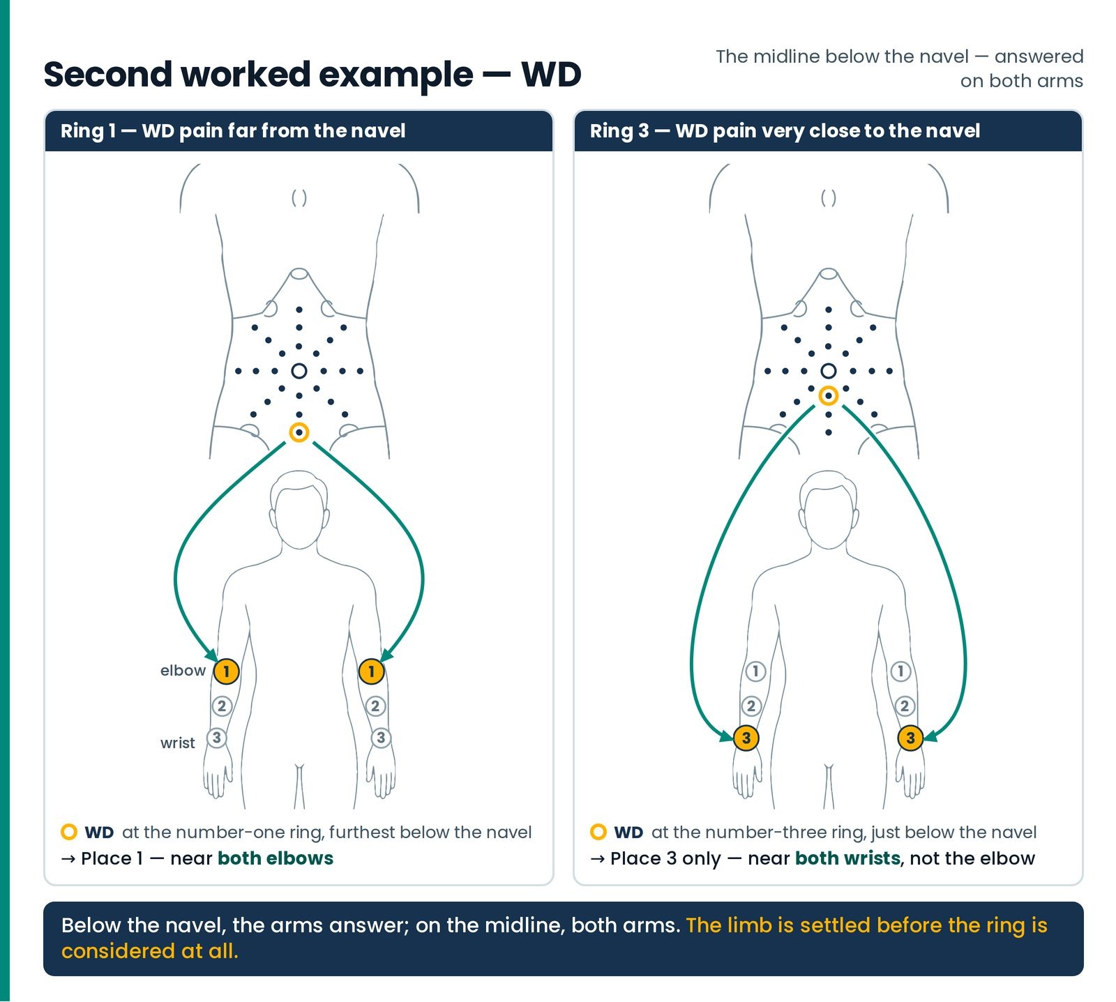
The pain nearest the navel will go only from the number-three treatment place.
Read that last line again, because it holds the surprise: the relationship is inverted. As the tender point moves inward towards the navel, the treatment site moves outward along the limb.
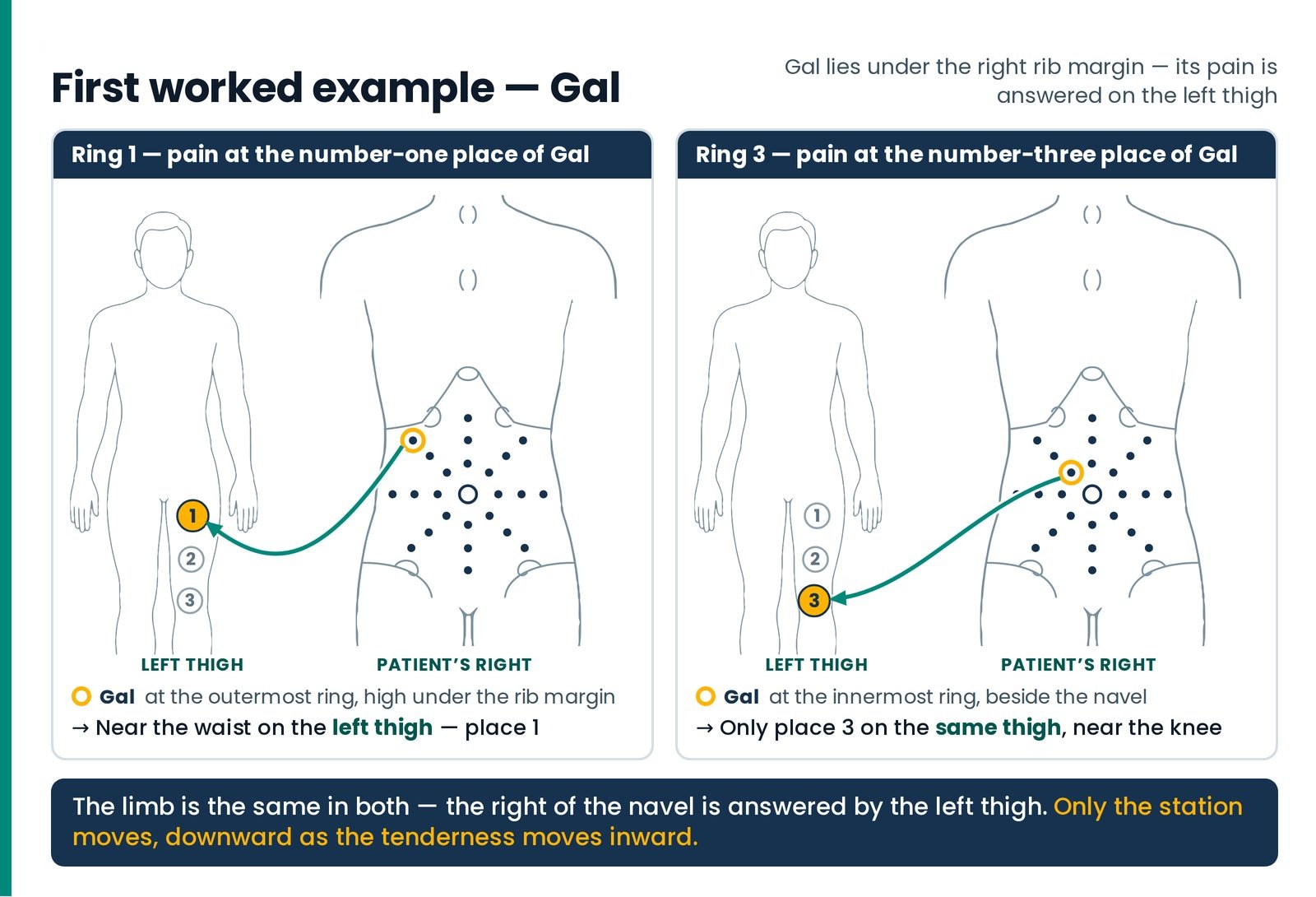
This is not an arbitrary convention to be memorised item by item but a single consistent transformation, which a student can apply to a point he has never seen before — a large part of why this system passes intact from teacher to student. Combine it with the four correspondences already taught — legs for pains above the navel, arms for pains between the navel and the perineum, left limbs for the right of the navel and right limbs for the left — and any square inch of the abdomen has an address on a limb.
The two worked examples
Two worked examples carry the rule, covering both limbs, both directions of the crossed rule and both ends of the ring. (Figure 6.4) (Figure 6.5)
Right follows left
Work with the bowel’s own wave — and the two sides of the sixth bead
One rule of order belongs to this article rather than the formulary, because it is a rule about the belly itself — and without it no student will understand why any formula is arranged as it is.
The peristaltic principle — right follows left
Pressure cannot be applied in any random sequence.
The general rule for proper peristaltic movement is: right follows left. That is — first stimulate the organs on the right side of the navel, and then stimulate the organs on the left side of the navel.
A reverse order can cause constipation, and is to be resorted to only when we wish to arrest the peristalsis — for example while treating loose motions.
Peristalsis is worth a sentence of plain English, because the whole rule hangs on it. The intestine is a long muscular tube that moves its contents along by squeezing behind and relaxing in front, in a travelling wave — the way you work a stiff toothpaste tube from the closed end. That wave has a fixed direction: up the right side of the abdomen, across the top, down the left, out. Right, then left. Everything in the belly runs that way.
So the ordering rule is not a ritual but a decision to work with the wave. Stimulate the right-hand organs first and the left after, and you have pushed in the direction the bowel was already going. Reverse it and you have pushed against it — which slows the bowel and, in a patient already sluggish, constipates him. And precisely because reversing it slows the bowel, reversing it deliberately is a treatment: for loose motions, arresting the wave is the object. That is why reversed forms of certain formulas exist, why they are given for a short time only, and why the normal order must be restored afterwards.
The same principle of sided opposites appears in a second place a therapist meets early, and the two teach one lesson.
The two sides at the sixth thoracic bead
Pressure applied on the left side of the spine, in the region of the sixth thoracic vertebra, in a particular fashion, has a dilatory function — it opens — and is used in situations associated with narrowing of arteries or of the ring-shaped closing muscles of the body: for example, narrowing of the food pipe. Left Prone
A similar pressure applied on the right side of the spine constricts — it closes — and has been used to relieve, effectively and in a great many patients, the swelling in the legs caused by varicose veins. Right Prone
Same bead. Same manner of pressure. Two sides. Opposite results.
Nothing makes the point about sides more sharply. Two treatments, an inch or two apart on the same back, and one opens what the other closes. A therapist vague about which side he worked has not been slightly imprecise; he has possibly given the opposite treatment.
What you are actually pressing over
Four fields of the trunk, and why a code name is not an address
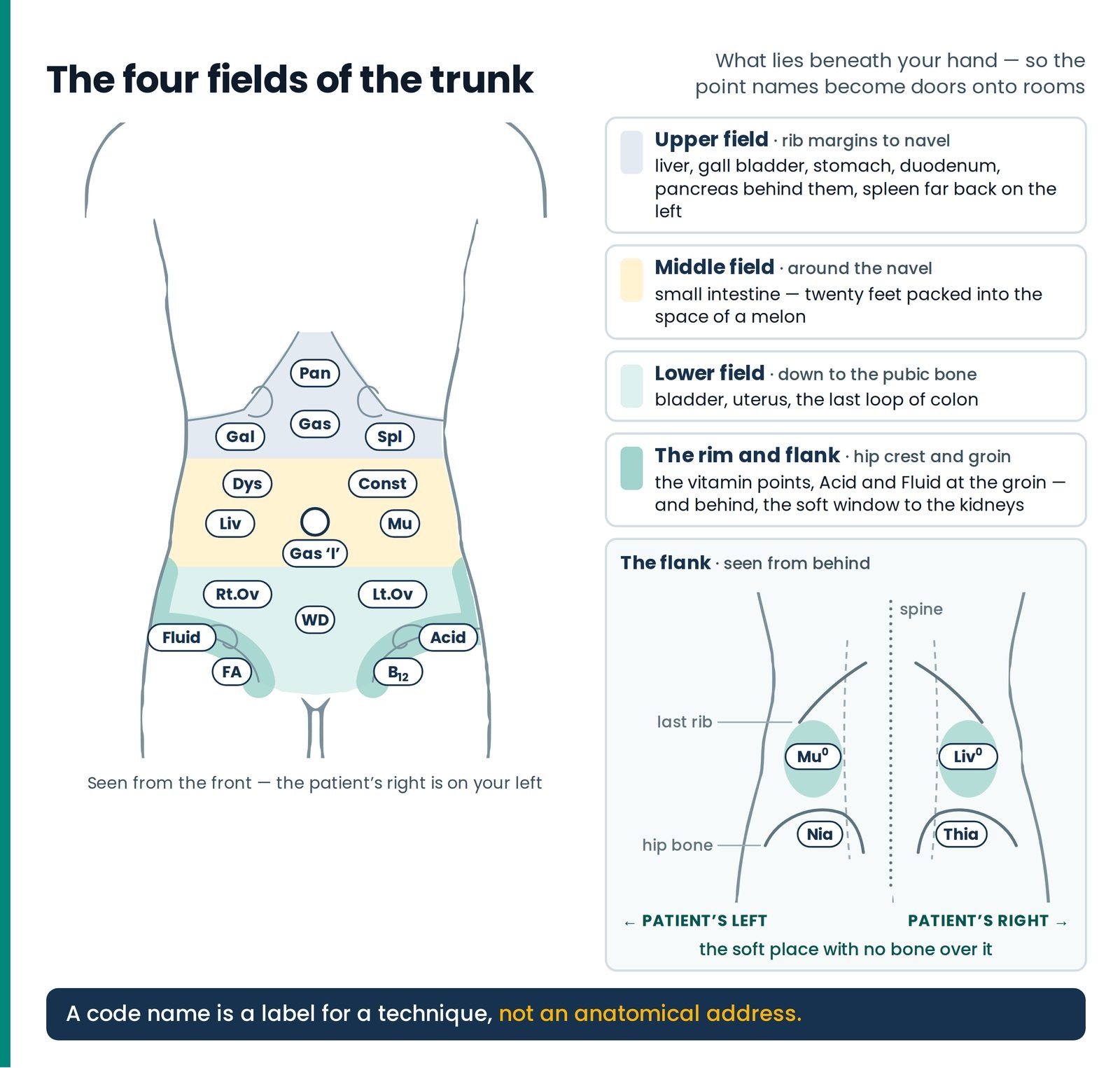
A therapist who knows what is beneath his hand presses differently, and stops at the right moment. Four fields hold the whole map: the upper field, rib margins to navel — liver, gall bladder, stomach, duodenum, pancreas behind them, spleen far back on the left; the middle field around the navel — small intestine, twenty feet packed into the space of a melon; the lower field down to the pubic bone — bladder, uterus, the last loop of colon; and the rim and flank, carrying the vitamin points, Acid and Fluid at the groin, and behind, the soft window to the kidneys. (Figure 6.7)
The code names are, the teaching itself says, in general indicative of the major organ of that area — though not necessarily always so, and the qualification is load-bearing. Spl is the clearest case: a healthy spleen sits high and far back on the left, behind ribs nine to eleven, and cannot be felt through a belly wall at all. Lt.Ov is another — an ovary lies deep on the side wall of the pelvis, and in a man there is no gonad in that quadrant. So the honest statement of the map is: tenderness at the place named Spl accompanies disorders connected with the spleen, and treating it helps. A claim about a finding and a technique, not a claim about what your fingertip is resting on. (Figure 6.6)
Two bodies, read from the navel
A camp lesson and a four-complaint case, read from the pain points
Questions students ask
1. I am twenty and I cannot feel anything at all. Every belly feels the same to me. Is my hand no good? Your hand is fine; your reference library is empty. A finding only means something in comparison, and you have not yet examined enough people to have anything to compare with. Examine willing family members at the same three points and write down what you find each time. Within a month the bellies will stop feeling the same. See also → The Educated Hand, "The Educated Hand".
- 2. I am a retired bank manager, sixty-six, learning this for my wife. Must I memorise all twenty names first? No. Learn six with their landmarks — Pan, Gal, Spl, Liv, Mu, Const — and the two flank points. Those eight cover most of what you will meet, and the rest attach themselves as you meet them, which is a better way of learning than a list.
3. I am a doctor. You call Spl the spleen point, but nobody can palpate a normal spleen there. What exactly is being claimed? Exactly what you have identified, and the teaching itself flags it: the names are in general indicative of the major organ of the area, though not necessarily always so. The claim is that tenderness at that site accompanies disorders connected with the spleen and that treating it helps — a claim about a finding and a technique, not about what your fingertip is resting on.
- 4. I work in a village, and my patients are labourers with very hard abdominal walls. The landmarks disappear. Use bone, not tissue. The bottom of the breastbone, the rib margins, the crest of the hip bone and the navel are there whatever the muscle is doing. Have the patient bend the knees up, which lets the wall go slack, and press on the out-breath.
- 5. I am a housewife and the number boxes confused me for weeks. Is a three worse than a one? No — and that mistake costs treatments. The number is a position, not a grade: three is nearest the navel, one furthest out. The answering station on the limb runs the other way, ring three being treated near the knee or the wrist.
- 6. If Liv is on the right of the navel, why is it treated on the left thigh? The crossed rule: pressure on the left limbs relieves pains on the right of the navel, and on the right limbs, pains on the left. Combine it with the upper-and-lower rule — legs for pains above the navel, arms for pains below — and any quadrant has an address. See also → The Hand That Heals, "The Hand That Heals".
- 7. I have no science background and I get lost the moment you say what is under the skin. How much must I know? Enough to press respectfully and to know when to stop. Under your finger is skin, then fat, then muscle, then a thin lining, and only then anything hollow or solid — and at the flank, in that one soft gap between last rib and hip bone, there is no bone over the kidney at all.
- 8. My teacher checks Mu° and Liv° before anything else. Why start at the back? Because that comparison sorts the patient into one of two great families before you have spent twenty minutes on anything else, and much of the plan follows from it. What the two flanks mean is The Two Kingdoms, "The Two Kingdoms".
- 9. A patient's belly is tender absolutely everywhere I press. What does that tell me? Most likely that you are pressing too hard — the first thing to exclude. Possibly that she is frightened. And possibly that you should not be pressing this abdomen at all: if a belly is rigid, exquisitely tender, or comes with vomiting and distension, stop and send.
Questions patients ask
- 1. "My problem is my knee. Why are you pressing my stomach?" Because the knee is repaired out of the same stores as everything else, and those stores come from the belly. Before I do anything to your knee I want to know how well your digestion is working, and the quickest honest way to find that out is with my fingers, here, in about two minutes. If I find nothing I will say so.
2. "There is pain there! I never knew there was pain there. Is something wrong inside me?" It is a common finding and it is not a frightening one. A tender spot there tells me that a part of your abdomen is not getting the blood flow it should — not that anything is damaged. Most people carry two or three of these without ever knowing, because nothing in ordinary life presses on them. It usually settles as we work, and you will notice it settling.
- 3. "Will you have to press hard?" No. Firm enough to feel something, soft enough not to make it. If I press hard enough to hurt anybody, I will find pain in everybody, and then the examination tells me nothing. If anything is uncomfortable, say so at once — I would rather know.
4. "What are those short words you write on my card? Is it something you don't want to tell me?" Not at all — ask me any of them and I will tell you. They are our shorthand for places on the body. This one, Liv°, is our word for the kidney. We use a short name because kidney is a word that frightens people out of all proportion, and there is nothing here to be frightened of. Nothing goes on that card that I would not say to you out loud.
5. "My doctor says my kidney reports are normal. Then why is this spot painful?" That is good news and worth having: a normal report means nothing the test measures is wrong. What I am finding is a tender place in the wall of your back, which in our system tells me about the flow of blood in that region rather than about damage. The two are not in conflict, and I would not want you to think your normal report is secretly abnormal — it is not.
6. "Can you tell what disease I have just by pressing?" No, and be careful of anyone who says they can. What this tells me is which regions of your abdomen are not working freely today. That is a great deal — it is what I build the treatment on — but it is not the same as naming a disease. For that, your doctor's tests do a job my hands cannot do, and I will tell you plainly when I think you need one.
Words used here
| Hindi | English | Neurotherapy code / usage |
|---|---|---|
| नाभि (nābhi) | Navel | The centre of the body; the reference point from which every front pain point is located |
| नाभी का खिसकना (nābhī kā khisaknā) | Slipping of the navel | The folk condition behind navel setting; called धरन (dharan) and पिछोटी (pichhoṭī) in Punjabi, डूटी (ḍūṭī) and बाइगोला (bāigolā) in UP and MP |
| दर्द के प्वाइंट (dard ke point) | Pain points | The pre-determined places at which tenderness or hardness is sought; sixteen on the front chart, four on the back |
| कमर की हड्डी (kamar kī haḍḍī) | Hip bone; the crest of the pelvis | The lower edge of the flank window, and the rim carrying the four vitamin points |
| पसली (paslī) | Rib | The last rib is the upper edge of the flank window for Mu° and Liv° |
| गुर्दा (gurdā) | Kidney | Never named to an anxious patient: Mu° for the left, Liv° for the right |
| जिगर / यकृत (jigar) · पित्ताशय (pittāśay) · तिल्ली (tillī) · अग्न्याशय (agnyāśay) | Liver · gall bladder · spleen · pancreas | Liv right of the navel · Gal under the right rib margin · Spl under the left · Pan at the pit of the stomach |
| आँव (āṃva) | Slimy mucus in the motions | The finding that names Mu; the code covers the wet lining of the whole digestive tract |
| कब्ज़ (kabz) · दस्त (dast) | Constipation · loose motions | Const left upper quadrant · Dys right upper quadrant, working against peristalsis |
| सफ़ेद पानी (safed pānī) | White discharge | WD — the midline below the navel; uterus and prostate |
| आँत की गति (āṃt kī gati) | The motion of the intestines — peristalsis | The wave that fixes the rule right follows left |
| — | Ring · renal angle | The number 1, 2 or 3 beside a point name is a distance from the navel, not a severity · the medical name for the same flank window in which Mu° and Liv° are found |