Watch


The back chart — Mu° and Liv°
The kidney points in the soft hollow between last rib and hip bone
Turn the patient, or reach round to the flank, and the map continues. The back list is short, and it is the half of the chart a therapist uses first.
Table 3 — The pain points of the back
| Code | Read aloud | Principal organ or related symptom | Where it lies |
|---|---|---|---|
| Mu° | Mucus zero | Left kidney | The soft part in the middle, between the last rib and the hip bone, on the left Left |
| Liv° | Liver zero | Right kidney | Exactly the same place on the right Right |
| Thia | Linked to thiamine deficiency symptoms | Above the back of the hip bone Right | |
| Nia | Linked to niacin deficiency symptoms | Above the back of the hip bone Left |
Finding Mu° and Liv° by feel
You find this point with your hand and not with a ruler, so it is taught by feel.
Why that spot is soft, and what sits behind it
The softness is not fat and not an accident. It is the one window into the back of the abdomen that no bone covers.
Going in: skin and fat; then the strong sheet of fascia across the low back into which the great back muscle inserts; then, in the small triangular gap between that muscle, the flat muscle of the flank and the rim of the hip bone, a place where the wall is genuinely thin — which is why your finger reports soft. Deeper, the square muscle running from hip rim to last rib, and beside it the great hip flexor; crossing between the layers, the nerves carrying sensation from this strip of skin — which is why tenderness here can be muscular or nerve-borne rather than anything to do with a kidney. Then the space behind the abdominal cavity proper: a membrane like a paper bag packed with fat, and in that fat the kidney, the right one about half a bone lower than the left because the liver takes the room above it. Capping each kidney like a small hat, the adrenal gland.
The two reasons for the code names
Now the naming, which is one of the most humane pieces of thinking in the whole therapy.
Why the kidneys are called Mu° and Liv°
The first reason is to spare the patient alarm.
Stimulation of the kidney is a necessary part of a great many treatments, so the word would otherwise be spoken constantly — across a treatment room between therapists, and in writing on a card the patient can read. But kidney is a much-maligned word: in many patients it conjures dialysis, hospital wards, an operation, and in an anxious one the mere utterance of it can send the blood pressure up. To save patients from such baseless fears, the kidneys were given the nicknames Mu° and Liv°.
The second reason is memory. The Liv pain point lies on the right of the navel, and the right kidney lies immediately behind it — so the right kidney is called Liv°. Right The Mu pain point lies on the left, and the left kidney sits just behind it — so the left kidney is aptly called Mu°. Left
Learn the two front points and you have the two back points for nothing. That is the whole design.
And the ring itself carries information. It is not decoration and it does not mean zero.
What the small ring means
The place and the manner of giving treatment for Liv and Liv°, and for Mu and Mu°, are the same. But there are two changes in the method, and the ring records both.
One — one place instead of three. Where Liv and Mu are given from three places in turn along the limb, the ° points are given at a single point; to indicate that, the sign ° is used.
Two — the hand point moves up to the shoulder. The site of the point on the arm is up on the shoulder, shown by writing the ring as a superscript: the mark sits high, above the line, to remind the therapist that the contact goes high, near the shoulder joint, and not at the elbow where Liv and Mu take it.
One mark, at once a warning, a memory aid and a picture of the instruction.
At every step you can see the thoughtfulness behind this: with two logically worded abbreviations, therapist and patient were both put at ease in one stroke.
Two rules keep that kindness honest. A code may conceal a word; it may never conceal a finding — if you find something that ought to send a patient to a doctor, the nickname stops there and you say the real thing plainly, to the patient and on the paper he carries. And when a patient asks what it means, tell him: "It is our shorthand for the kidney. We use a short name because the word frightens people, and there is nothing here to be frightened of." A patient who discovers later that you had a private word for his kidney will not remember it as tact. ▲ Take care
What a finding at Mu° or Liv° means belongs to the next stage of your training. Tenderness at these two is the first thing checked in a Neurotherapy examination, and it sorts the patient into one of two great families — the acid direction or the alkaline — from which a very large part of the plan follows. That is taught completely in The Two Kingdoms, "The Two Kingdoms", with the two-column symptom picture, the dilution rule and the asymmetry of the two kidneys. Learn the anatomy and the naming here; learn the reading there.
The back pain chart
Twelve sites where the back reports an internal organ’s trouble
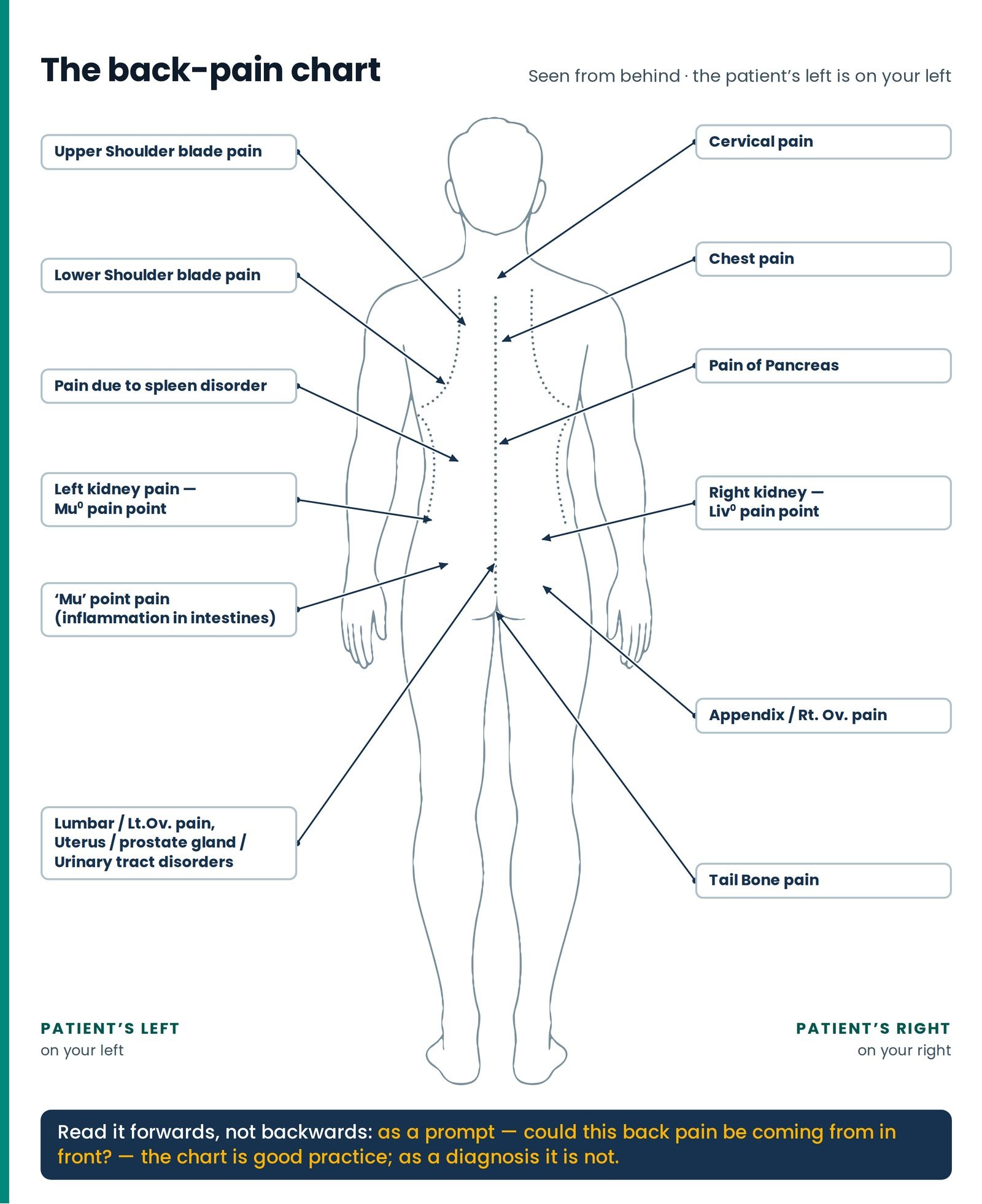
The second full-page chart is a whole-body outline seen from behind, head to heels, the spine a dotted line down the middle, the shoulder blades and lower rib margins dotted curves either side. Twelve labelled call-outs run around it, and together they make a compact statement of something the body does constantly and confusingly: it reports the trouble of an internal organ as an ache in the back wall. (Figure 6.2)
One drafting point must not catch you. The drawing is a view from behind, so the patient's left is on the reader's right — but the twelve call-out boxes have been placed for the sake of the page, the left-sided labels sitting on the reader's left and the right-sided ones on his right, the arrangement that would be correct facing the patient's front. A student who learns the chart as a picture will reverse every side on it the first time he stands behind a patient. Locate these points by anatomy, never by the side of the page: spleen and Mu° on the patient's left; liver, Liv° and appendix on the patient's right. Left Right
Table 4 — The back pain chart, label by label
| Label on the chart | Where the arrow lands | What lies at that level |
|---|---|---|
| Cervical pain | Nape, base of the neck, midline | The seven neck bones and the long muscles either side |
| Upper Shoulder blade pain | Upper scapular region, inside the shoulder blade | Upper trapezius, the muscle that lifts the shoulder blade, the small rhomboid, over ribs two to four |
| Lower Shoulder blade pain | At the bottom corner of the shoulder blade | Large rhomboid, lower trapezius, ribs six to eight; the base of the lung deep to it |
| Chest pain | Upper back, beside the spine | The first six thoracic beads, the long back muscles, and the joints where each rib meets the spine |
| Pain due to spleen disorder | Lower thoracic, below the shoulder blade | Ribs nine to eleven and the fascia of the low back; deep to them, spleen left, liver right |
| Pain of Pancreas | Mid-thoracic beside the spine | Long back muscles, ribs ten to twelve; the pancreas lies at the front of this level |
| Left kidney pain — Mu° pain point | The flank, between the twelfth rib and the rim of the hip bone Left | The square muscle of the low back, the hip flexor, the fat around the kidney, and the kidney |
| Right kidney — Liv° pain point | The mirror-image flank Right | The same, the right kidney about half a bone lower because of the liver above it |
| Mu point pain — inflammation in intestines | Lower lumbar, out from the spine | Long back muscles and fascia; deep and in front, the descending colon and small-bowel loops |
| Lumbar / Lt.Ov pain — uterus, prostate gland, urinary tract disorders | Lower lumbar and upper sacral region | The junction of low back and sacrum, the joints between sacrum and hip bones; deep in front, the tubes from the kidneys, the bladder, the uterus or prostate |
| Appendix / Rt.Ov pain | Lower lumbar to the sacro-iliac joint, opposite side | That joint and the bony knob behind it; deep in front, the caecum and appendix, or the pocket in which the ovary sits |
| Tail Bone pain | Coccyx, top of the cleft of the buttocks | The tail bone and its joint with the sacrum; the muscles of the pelvic floor taking their attachment there |
The chart is intelligent about a real thing. A pancreas in trouble really does bore through to the mid-back and ease when the patient leans forward; a kidney in trouble really is felt in the loin between last rib and hip rim, running towards the groin as a stone travels; trouble in the womb or prostate really is felt low in the back and over the sacrum. But read it forwards, not backwards. Most back pain at every one of these levels is muscle and posture, and an ache between the shoulder blades is far more often a woman who sews for nine hours than a pancreas. As a prompt — could this mid-back pain be coming from in front? — the chart is good practice; as a diagnosis it is not.